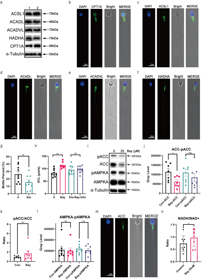
Fig. 6. IKBA phosphorylation inhibition by Bay117082 regulates FAO in human sperm.
a Expression of ACSL1, ACADL, ACADVL, HADHA, and CPT1A in sperm. α-Tubulin was applied to assess protein loading. Numbers represent samples of different individuals. b–f ACSL1, ACADL, ACADVL, HADHA and CPT1A localization in sperm. DAPI (blue) labeled the nuclei. CPT1A (b), ACSL1 (c), ACADL (d), ACADVL (e), and HADHA (f) were stained with AlexaFluor488 (green). Images were merged (MERGE) with bright-field images (Bright). Scale bar: 5 μm. g Motile percent of sperm treated with etomoxir (400 μM) for 1 h in BWW media. Values represent mean ± SEM (n = 9), **p < 0.01, compared with the corresponding control (0 μM). h Changes in VCL of sperm treated with 25 μM Bay117082 in the absence or presence of etomoxir (400 μM) for 1 h. Values represent mean ± SEM (n = 9), **p < 0.01, compared with the corresponding control. ns, no significance. i–l Western blotting showing AMPKA, ACC, pAMPKA, and pACC levels (i) in sperm treated with or without Bay117082 at 25 μM for 10 min. Gray-scale value analysis of ACC and pACC (j), pACC/ACC (k), as well as AMPKA and pAMPKA (l). n = 7, *p < 0.05, compared with the corresponding control (0 μM). ns, no significance. α-Tubulin was applied to assess protein loading. m Indirect immunofluorescence showing ACC localization in human sperm. DAPI (blue) labeled the nuclei; ACC was stained with Alexa Fluor 488 (green). Images were merged (MERGE) with bright-field images (Bright). Scale bar: 5 μm. n Changes in NADH/NAD+ levels in sperm treated with or without Bay117082 in BWW-GPL media. Values represent mean ± SEM (n = 8), *p < 0.05.

