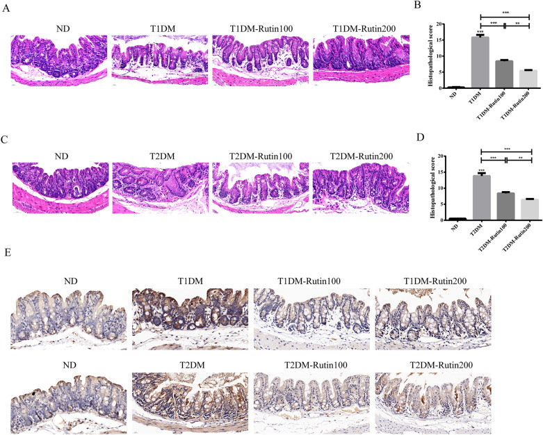
Figure 3.
Rutin improved colon lesions in diabetic mice. (A) H&E staining of colon tissues in ND, T1DM, T1DM-Rutin100 and T1DM-Rutin200 mouse groups (scale bar: 20 μm). Red arrows: crypts; black arrows: villi. (B) Histopathological scores of colon tissues in T1DM mice using H&E staining. (C) H&E staining of colon tissues in ND, T2DM, T2DM-Rutin100 and T2DM-Rutin200 groups. Red arrows: vacuole; black arrows: villi (scale bar: 20 μm). (D) Histopathological scores of colon tissues in T2DM mice by H&E staining. (E) Immunohistochemistry indicated that collagen I protein levels were increased in diabetic mice, which was recovered by rutin treatment (scale bar: 20 μm). Values are means ± SD (n = 5): *p < 0.05, **p < 0.01, ***p < 0.001.

