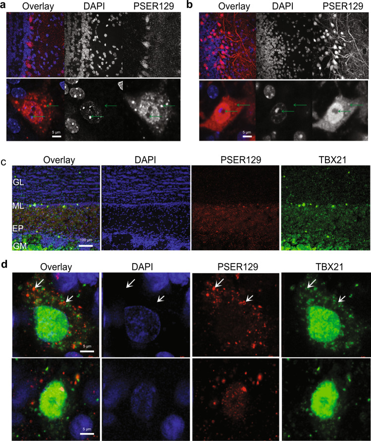
Fig. 2. Localization and intracellular distribution of PSER129 in the non-diseased OB.
a PSER129 was fluorescently labeled using TSA in WT mice. Tissues were counterstained with DAPI. Representative images showing the distribution of PSER129 in the OB. b Non-pathology bearing M83 mice labeled for PSER129. Non-human primate OB was dual-labeled for PSER129 and mitral cell marker TBX21. c Low magnification confocal images showing regional distribution in non-human primate OB. d High-magnification confocal images showing PSER129 puncta in TBX21-positive mitral cell. Green arrows denote intracellular punctate structures, both perinuclear and nuclear. WT mice n = 5, M83 mice n = 2, non-human primates = 2.

