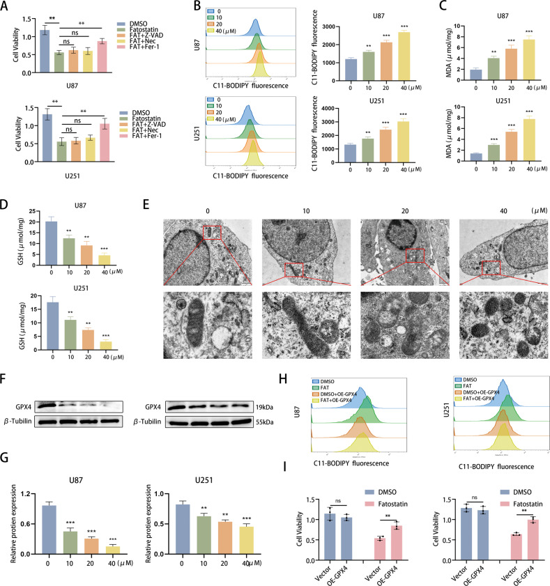
Fig. 4. Fatostatin induces ferroptosis mediated by GPX4 in GBM cells.
A According to the cell viability assay, Fer-1 (10 µM) reversed the decreased cell viability (absorbance value at 450 nm) of U87 and U251 cells induced by fatostatin, while Z-VAD (20 µM) and Nec (20 µM) did not. Z-VAD (Z-VAD-FMK), Fer-1 ferrostatin-1, Nec necrosulfonamide. B Flow cytometry was used to detect lipid ROS in U87 and U251 cells after fatostatin treatment. C, D MDA and GSH levels in U87 and U251 cells after fatostatin treatment were detected. E After DMSO and fatostatin treatment (20 µM), U87 cells were prepared for transmission electron microscopy observation. F, G The expression level of GPX4 in U87 and U251 cells after fatostatin treatment for 24 h. H Flow cytometry results showed that overexpression of GPX4 reversed the lipid ROS level in U87 and U251 cells. I CCK-8 assays showed that overexpression of GPX4 preserved cell viability (absorbance value at 450 nm) in U87 and U251 cells upon fatostatin treatment. *P < 0.05, **P < 0.01, ***P < 0.001; ns no significance.

