Figure 3.

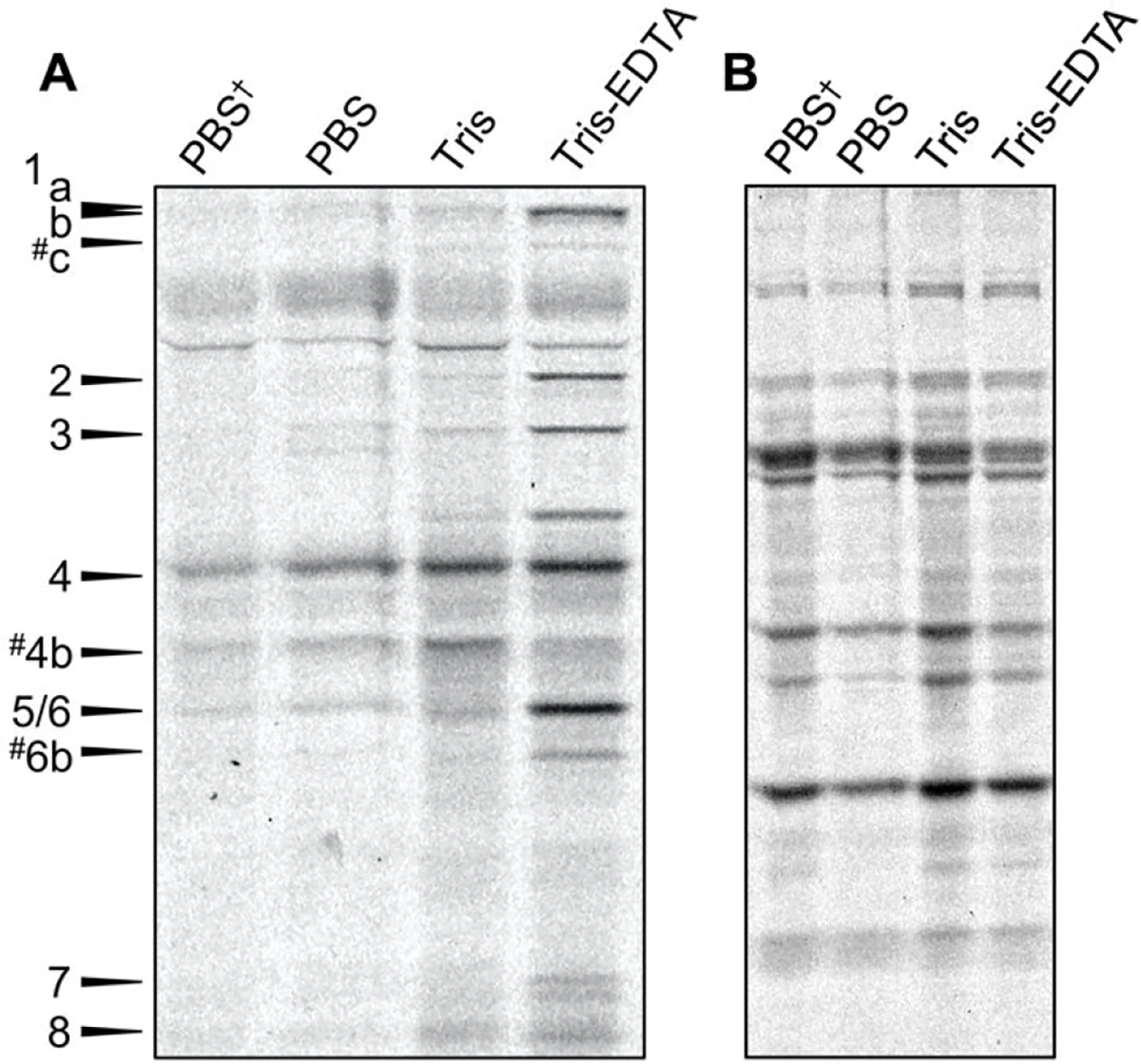
(A) Comparison of Bocillin-FL labeling using PBS, Tris, or Tris-EDTA. SDS-PAGE analysis revealed that treatment of E. coli using 25 μM Bocillin-FL in 50 mM Tris–200 μM EDTA for 30 min results in labeling of 11 or 12 PBPs in E. coli. (B). A Coomassie stain of the same gel indicates that the amount of proteome loaded into each lane is equivalent. PBS† indicates the previous DC2 procedure (7.5 μM Bocillin-FL in PBS, 10 min). # indicates putative PBP assignments. One of three biological replicates shown.
