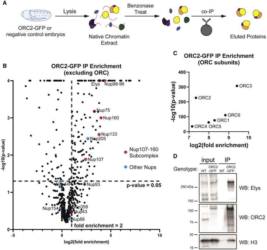
Figure 1. ORC interacts with subunits of the nuclear pore complex.
(A) Schematic of extract preparation and immunoprecipitation protocol using ORC2-GFP or Oregon R (negative control) embryos.
(B) Average fold enrichment and statistical significance for three biological replicates of GFP-Trap immunoprecipitation (IP) mass spectrometry for ORC2-GFP-expressing embryos relative to negative control embryos. Highlighted are all nucleoporin proteins identified by mass spectrometry. Dashed lines indicate significant level cutoffs (<0.05 for p value and ≥2-fold enrichment).
(C) Same as (B) but with only ORC subunits.
(D) Western blots using anti-ORC2, anti-Elys, or anti-histone H3 antibody on samples derived from the IP.

