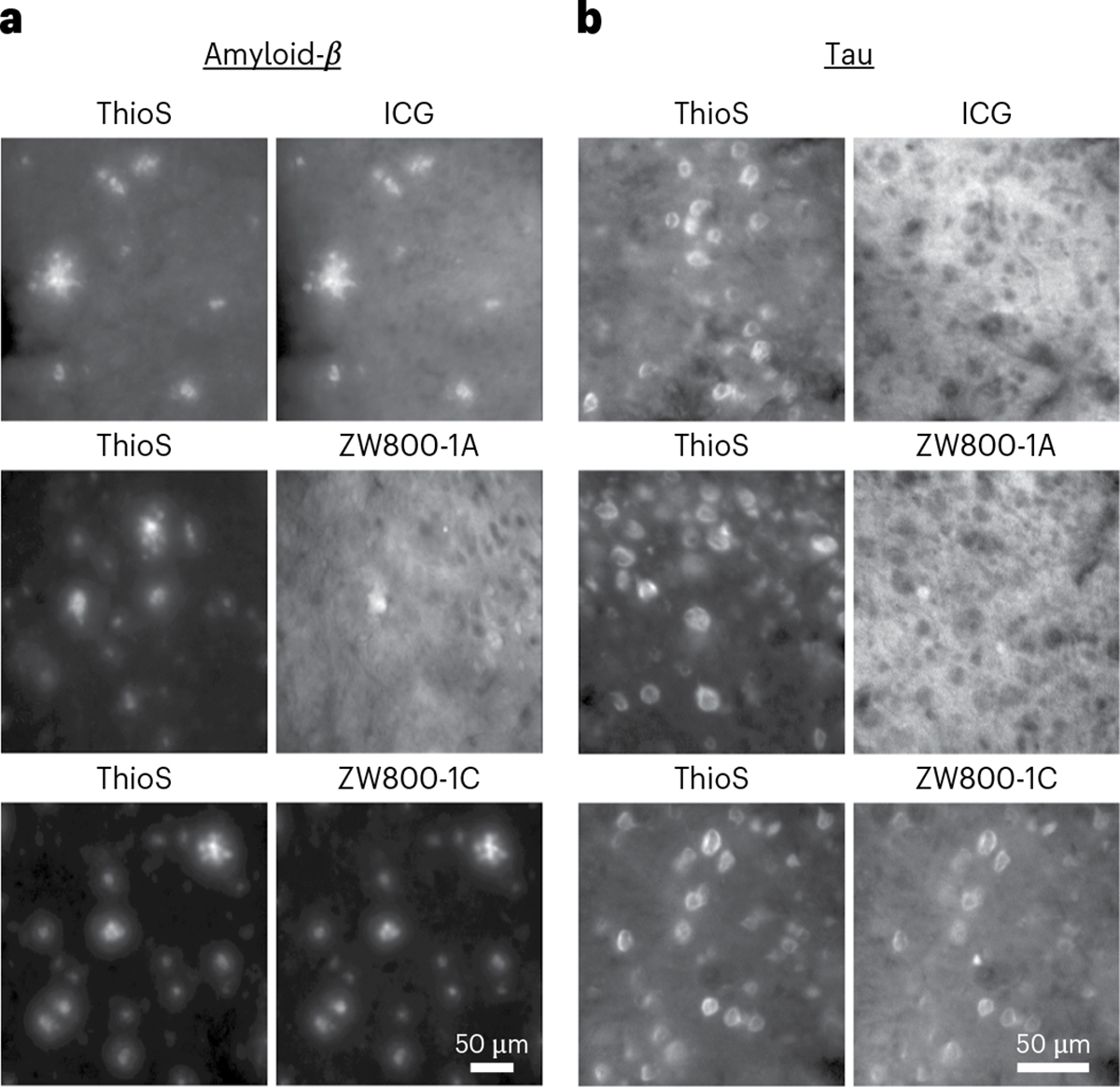
Fig. 2 |. Ex vivo imaging of amyloid plaques and NFTs in APP/PS1 and rTg4510 mouse brain sections.

a,b, APP/PS1 (a) and rTg4510 (b) mouse brain tissue sections co-stained with ThioS (0.05%) and each NIR fluorophore (100 μM). Images were acquired with widefield epifluorescence imaging in the green (for ThioS) and NIR channels.
