
Fig. 6 |. Two-photon microscopy and non-invasive lifetime imaging of AD pathology with ZW800–1C.
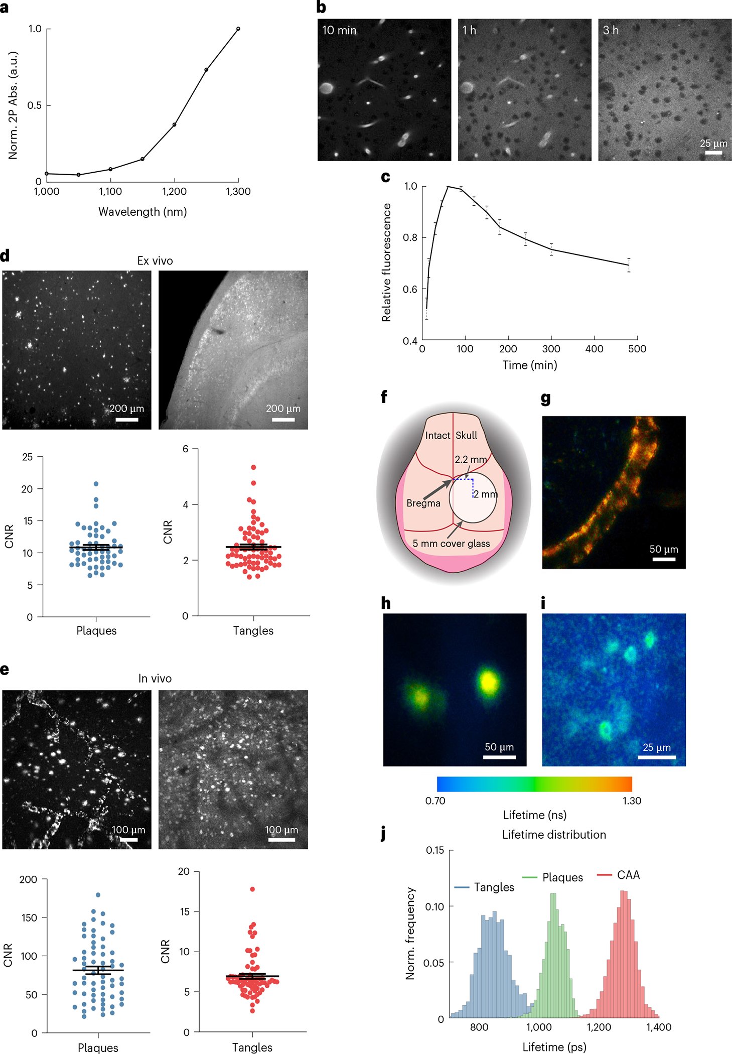
a, Two-photon absorption spectrum of ZW800–1C. b, Time-lapse images of ZW800–1C fluorescence in brain parenchyma of C57BL/6J mice after intravenous injection. c, Clearance dynamics of ZW800–1C out of the brain from t = 10 min to t = 8 h from n = 3 C57BL/6J mice. d,e, Quantitative CNR analysis comparing ex vivo (d) and in vivo (e) detection of amyloid plaques and neurofibrillary tangles using two-photon microscopy. Top panels show representative images while bottom panels show CNR of individual plaques and tangles. f, Schematic showing the region on the intact skull for non-invasive through-the-skull imaging. g–i, Representative images from n = 3 APP/PS1 Tg and n = 3 rTg4510 Tg mice demonstrating non-invasive lifetime imaging of CAA (g), amyloid plaques (h) and neurofibrillary tangles (i) through the intact skull using ZW800–1C (100 nmol). j, Histogram distribution of fluorescence lifetimes acquired through the intact skull after binding to AD pathology. Data are shown as mean ± s.e.m.