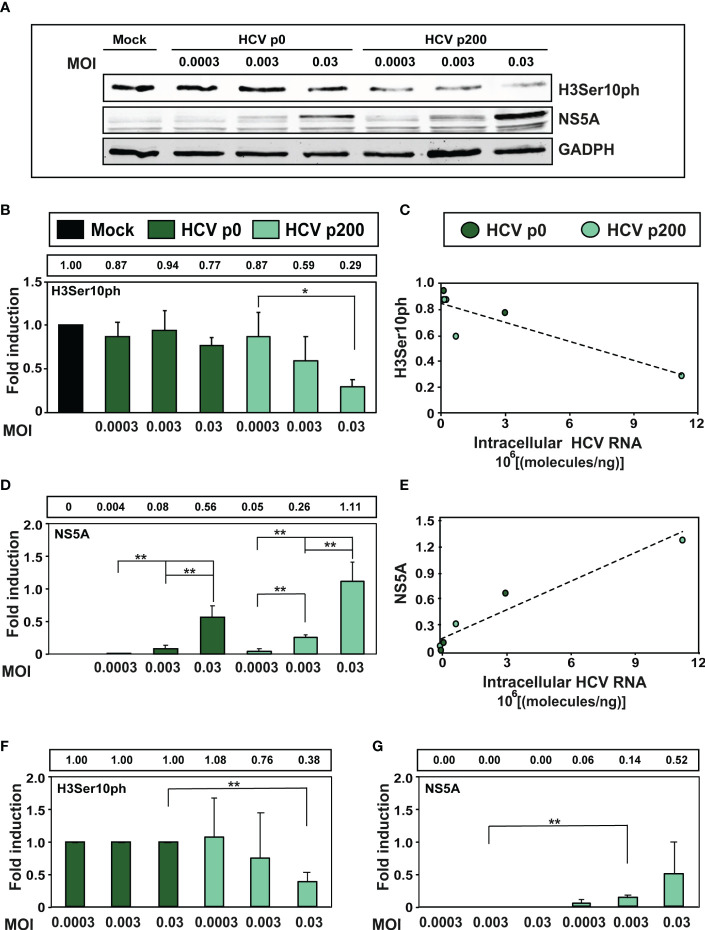
Figure 3.
Effect of the multiplicity of infection (MOI) on the intracellular H3Ser10ph level. (A) Western blot analysis of extracts of cells that were either mock-infected or infected with HCV p0 or HCV p200 at the indicated MOI. Extracts were prepared at 72h post-infection. GAPDH was used as the loading control. (B) H3Ser10ph levels in mock-infected or HCV-infected cells, expressed as the fold induction relative to the corresponding value for the mock-infected cells. The values are the average (and standard deviations) of the densitometric quantifications of three independent western blots; the image of one of them is shown in (A) The MOI and infecting virus are indicated in abscissa, and the densitometry values (using as reference the corresponding value for the extract of mock-infected cells, taken as 1) are written in the box above the panel. (C) Correlation between the intracellular amount of viral RNA measured at 72h post-infection (abscissa) and the H3Ser10ph level determined by densitometry of the Western blots (ordinate). The discontinuous line corresponds to function y=-5x10-0.8x+0.85 (R2 = 0.78; p-value=0.02027, linear regression test). (D) Same as B but for viral protein NS5A. (E) Same as C but for viral protein NS5A. The discontinuous line corresponds to function y=1x10-0.7x + 0.12 (R2 = 0.918; p-value=0.0017, linear regression test). (F, G) Same as B and D but with the H3Ser10ph and NS5A levels in HCV-infected cells expressed as the fold induction relative to the corresponding value for the HCV p0 infected cells. The statistical significance of differences is as follows: *p<0.05; **p<0.01; unpaired t -test.

