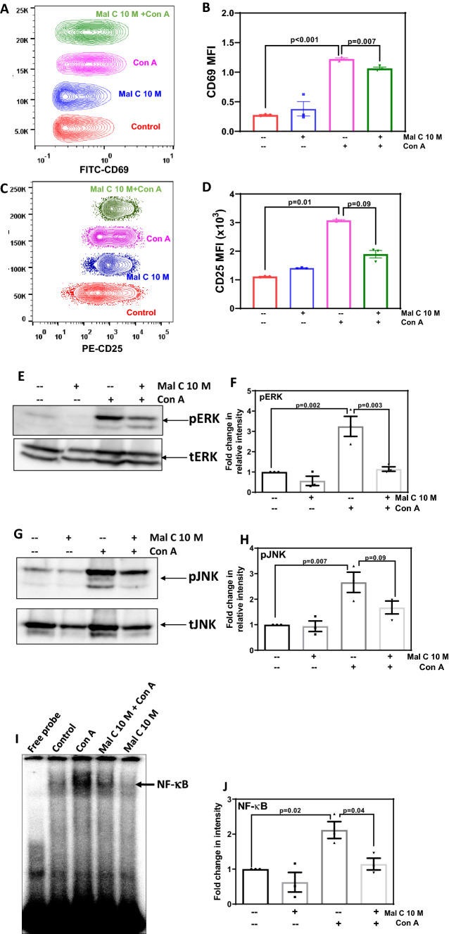
Figure 2.
Mal C suppressed the expression of T-cell activation markers, mitogen-induced phosphorylation of MAPK, and NF-κB DNA binding: Lymphocytes (2.5×10⁶) incubated with Mal C (10 μM) followed by stimulation with Con A (2.5 μg/mL) were subsequently cultured for 24 h at 37°C and then stained with anti-CD69-FITC (A and B) or anti-CD25-PE (C and D) conjugated antibodies. Lymphocytes stained in such manner were then acquired using a flow cytometer, and concatenated flow cytometric histograms are shown. The percentage of positive cells are graphed in (B) and (D). Three independent experiments were performed with data points representing mean±SEM from three replicates from one such experiment. Lymphocytes incubated with vehicle or Mal C (1 h) were treated with Con A (2.5 μg/mL for 3 h) followed by probing for pERK and ERK (E and F), and pJNK and JNK (G and H) in the whole-cell lysates by western blotting. The bar graphs show quantification of blots from three independent experiments (F and H). Nuclear extracts were prepared from lymphocytes incubated with Mal C 10 μM for 1 h followed by stimulation with Con A (2.5 μg/mL) for 4 h and subsequently probed for NF-κB DNA binding by EMSA (I). The bar graph in (J) shows the quantitation of NF-κB band intensity from three independent experiments.

