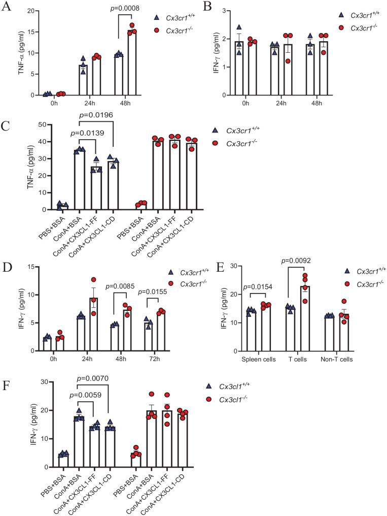
Figure 4.
Disruption of CX3CL1/CX3CR1 axis leads to enhanced pro-inflammatory properties of macrophages and T cells.
(A, B) TNF-α and IFN-γ levels in the supernatant of ConA-treated Cx3cr1+/+ and Cx3cr1−/− BMDMs (10 μg/mL). (C) BMDMs were pretreated with 100 ng/mL full-length CX3CL1 (CX3CL1-FF), 100 ng/mL the domain of CX3CL1 (CX3CL1-CD) or 100 ng/mL BSA, then incubated with ConA (10 μg/mL) for 48 h to detect TNF-α level. (D) IFN-γ levels of ConA-treated spleen cells (10 μg/mL) were detected by ELISA assay. (E) T cells and non-T cells in spleen cells were separated by pan T cells isolation kit, then total spleen cells, T cells, and non-T cells incubated with ConA for 48 h. IFN-γ levels of each group were measured by ELISA assay. (F) IFN-γ levels of the supernatant of T cells were detected after co-incubation with ConA + BSA, ConA + CX3CL1-CD and ConA + CX3CL1-FF. (A color version of this figure is available in the online journal.)

