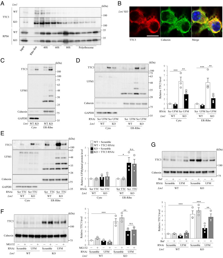
Fig. 2.
TTC3 is localized in 40S subunits and partially stabilized by UFMylation signal pathway. (A) Ribosome-free (Free), 40S, 60S, 80S monosome and polysome fractions were isolated from cultured cortical neurons by sucrose gradient centrifugation, and TTC3 and RPS6 (control) proteins were detected by western blotting. (B) Endogenous TTC3 was stained with an anti-TTC3 antibody (red), and ER was visualized with an anti-calnexin antibody (green). (Scale bar, 25 μm.) (C) Cytosolic (Cyto) and ER-associated ribosome-enriched (ER-Ribo) fractions were isolated from cultured cortical neurons by the centrifugation method and subjected to western blotting with an anti-TTC3 antibody. Calnexin was detected as an ER-bound ribosome-enriched associated membrane (ER-Ribo) fraction marker, and GAPDH was used as a cytosolic marker. (D) Cultured cortical neurons were infected with lentivirus encoding scramble RNAi or UFM1 RNAi, and cytosolic (Cyto) and ER-bound ribosome-enriched associated membrane (ER-Ribo) fractions were isolated. The signal intensities of TTC3 protein were normalized to those of GAPDH for cytosolic fraction or to calnexin for ER-associated ribosome-enriched fraction and then expressed as the relative ratio of WT neurons infected with scramble RNAi (Right) [Cyto: n = 3, F(3, 6) = 43.7, P = 0.0017; ER-Ribo: n = 4, F(3, 9) = 21.9, P = 0,0002, one-way ANOVA; **P < 0.01, ***P < 0.001, Bonferroni’s multiple comparison test post hoc]. (E) Cultured cortical neurons were infected with lentivirus encoding scramble RNAi or TTC3 RNAi, and cytosolic (Cyto) and ER-bound ribosome-enriched associated membrane (ER-Ribo) fractions were isolated. TTC3 and UFMylated proteins were detected by western blotting. The signal intensities of TTC3 protein were normalized to those of GAPDH for cytosolic fraction or calnexin for ER-Ribofraction and then expressed as the relative ratio of WT neurons (Right). [n = 3, Cyto: F(3, 6) = 1.59, P = 0.2877; ER-Ribo: F(3, 6) = 10.1, P = 0.0093, one-way ANOVA; *P < 0.05, Bonferroni’s multiple comparison test post hoc]. (F) Cultured cortical neurons with UFM1 KD were treated with or without 20 μM MG132 for 6 h, and ER-Ribo fraction was isolated. TTC3 was detected by western blotting. The signal intensities of TTC3 protein were normalized to those of calnexin and then expressed as the relative ratio of WT neurons infected with scramble RNAi and without MG132 treatment (Right). [n = 3, F(7, 14) = 41.7, P < 0.0001, one-way ANOVA; **P < 0.01, ***P < 0.001, Bonferroni’s multiple comparison test post hoc]. (G) Cultured cortical neurons infected with indicated lentivirus were treated with or without 200 nM bafilomycin A (Baf) for 6 h, and ER-Ribo fraction was isolated. The signal intensities of TTC3 protein were normalized to those of calnexin and then expressed as the relative ratio of WT neurons infected with scramble RNAi, without Baf treatment (Right) [n = 3, F(7, 14) = 43.7, P < 0.0001; one-way ANOVA; **P < 0.01, ***P < 0.001, Bonferroni’s multiple comparison test post hoc]. n.s., not significant. Throughout the figures, data represent means ± SEM.

