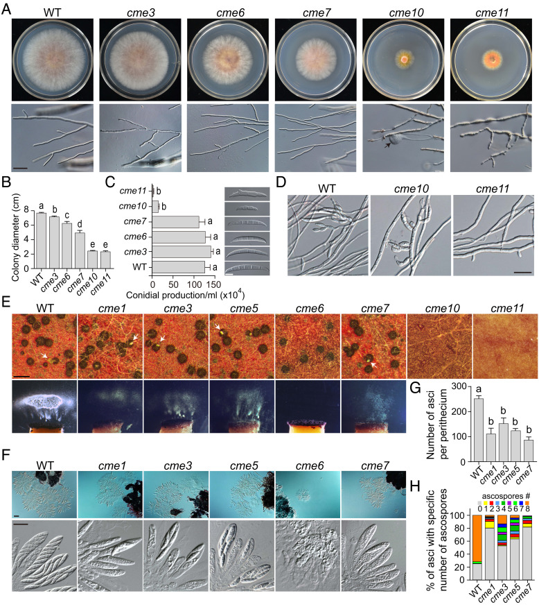
Fig. 2.
Defects of deletion mutants in vegetative growth, conidiation, and sexual reproduction. (A) Three-day-old PDA cultures and 48-h hyphal tips of PH-1 (WT) and the five marked deletion mutants. The black arrow points to the swollen tip of hyphae. Bar = 20 μm. (B) Colony diameters of 3-d-old PDA cultures of the marked strains. (C) Conidial production of the marked strains was examined by differential interference contrast (DIC) microscopy. Bar = 10 μm. (D) Phialides produced by the marked strains in 72-h carboxymethylcellulose (CMC) cultures were examined by DIC microscopy. Bar = 50 μm. (E) Mating cultures of the marked strains were examined for perithecium formation and ascospore discharge at 8 days postfertilization (dpf). White arrows indicate the ascospore cirrhi. Bar = 0.5 mm. (F) Ascus and ascospore formation of the marked strains were examined by DIC microscopy at 5 dpf (Upper, bar = 50 μm) and 8 dpf (Lower, bar = 20 μm), respectively. (G) The number of asci per perithecium produced by the marked strains at 5 dpf. (H) The percentage of asci containing a specific number of ascospores produced by the marked strains at 8 dpf. For (B), (C), and (G), mean and SD were calculated with data from three independent repeats (n = 3). Different letters indicate significant differences based on one-way ANOVA followed by Turkey’s multiple range test (P < 0.05).

