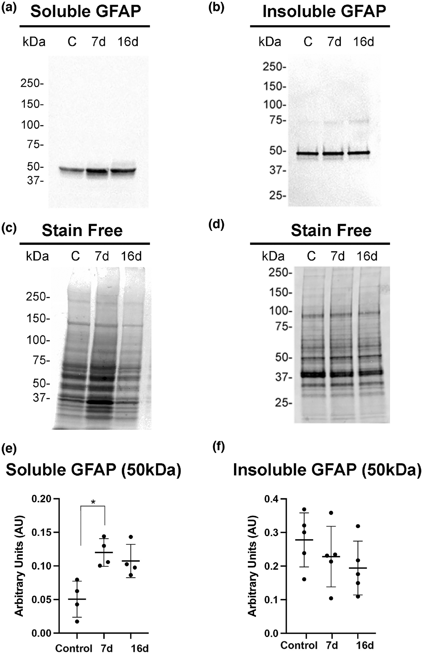
FIGURE 3.

Soluble GFAP increases in mouse retinas after laser injury. Soluble (a) and insoluble (b) cytoskeletal fraction were isolated from control uninjured and injured retinas (7- and 16-day post-injury) and subjected to western blot analysis. Blots were probed with GFAP antibody. (c,d) Stain-free gels were used as protein loading controls. Quantitation of GFAP bands normalized to stain-free gels was done using ImageJ software and data analyzed using GraphPad Prism software. (e) The Kruskal–Wallis test with Dunn’s multiple comparisons revealed a statistically significant difference of soluble GFAP abundance between uninjured and 7-day injured samples, H (2, N = 12) = 8.0; *p = .0121, but the 16-day injured sample did not show significant differences (p = .0997) (N = 10 mice/group). (f) The Kruskal–Wallis test with Dunn’s multiple comparisons did not reveal statistically significant differences for cytoskeletal GFAP abundance between uninjured and 7-day or 16-day injured samples, H (2, N = 15) = 2.94; p = .256. Data are presented as mean ± SD, p < .05 is considered significant.
