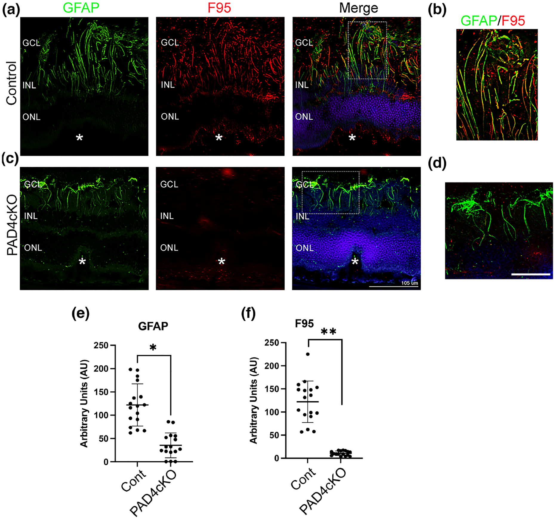
FIGURE 6.

Conditional knockout of PAD4 attenuates citrullination in mice subjected to retinal laser injury. Representative images from control (a) and PAD4cKO (c) retinas at 30 days post-laser injury. Cryosections were immunostained for GFAP (green) and citrullination with F95 (red). (a) F95 staining was evident on GFAP filaments in the lesions of control mice, while no F95 staining was observed in the lesions of PAD4cKO mouse retinas (c). Filamentous GFAP staining in PAD4cKO mouse retinas was reduced and sparse. (b,d) Enlarged images of the overlapping staining (Merge) boxed regions. The asterisks (a,c) denote the site of the laser lesion. GCL, ganglion cell layer; INL, inner nuclear layer, ONL, outer nuclear layer. Scale bar: a = 105 μm; scale bar: b = 35 μm. Quantification of GFAP (e) and F95 (f) staining using ImageJ software was performed (see File S3) and data analyzed using Mann–Whitney two-tailed nonparametric U test. Median GFAP staining of control samples and PAD4cKO samples were 120.0 and 29.75, respectively; the two groups were significantly different (U = 8, n1 = 17, n2 = 16, **p < .0001). Median F95 staining of control samples and PAD4cKO samples was 131.5 and 9.821, respectively; the two groups were also significantly different (U = 0, n1 = 17, n2 = 16, **p < .0001). N = 6 mice/group, average of three sections per mouse. Data are presented as mean ± SD, p < .05 is considered significant.
