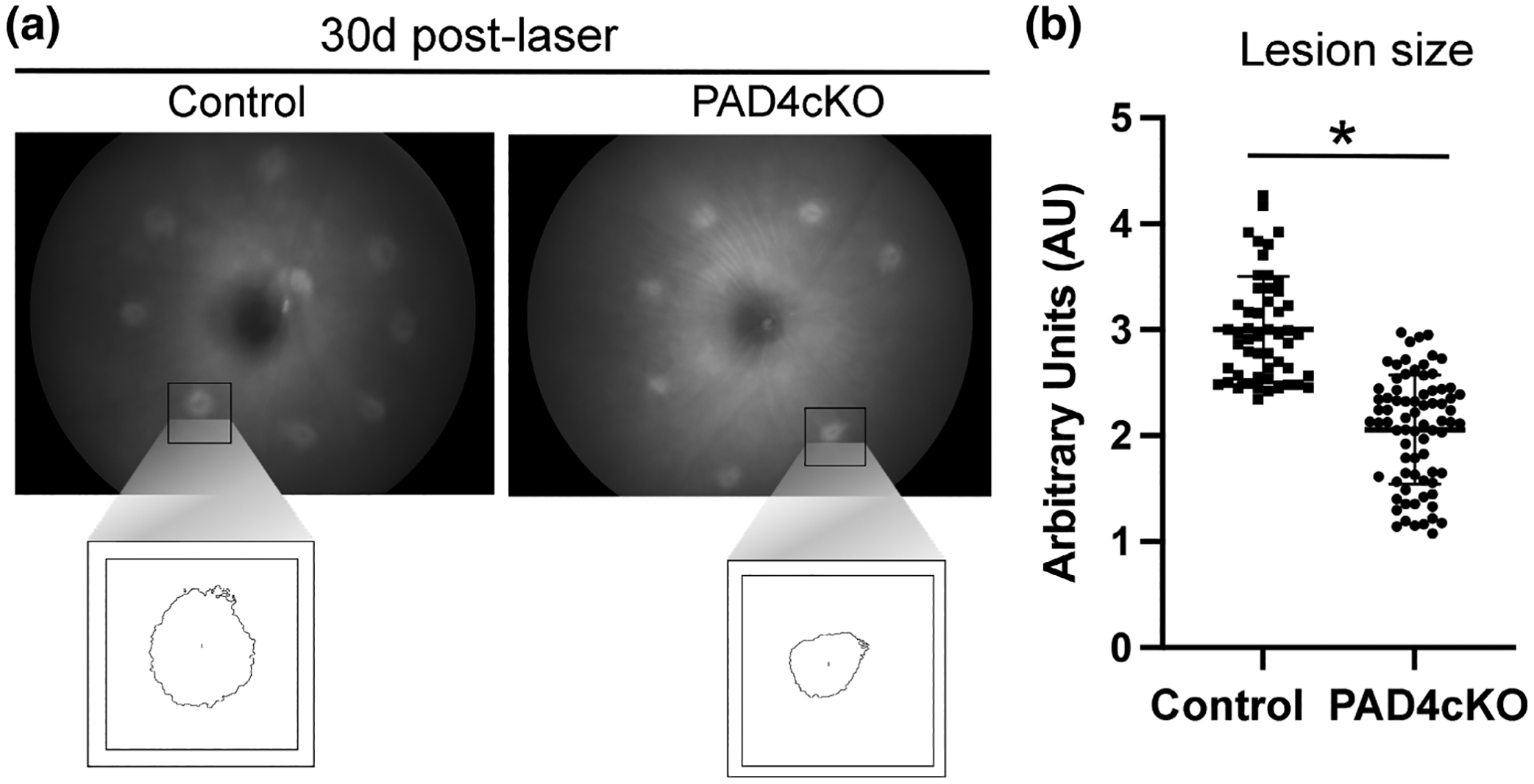
FIGURE 7.

Conditional knockout of PAD4 reduces lesion size of laser-damaged retinas. Retinal fundus images from control and PAD4cKO retinas from 30-day post-injured mice were acquired on a Phoenix Micron III fundus microscope. (a) Control mice revealed larger lesions compared to those from PAD4cKO mice. The boxed images represent measurements of individual lesions used for quantification. (b) Lesion sizes were quantified using ImageJ software and statistical analysis done using Mann–Whitney two-tailed nonparametric test. Median lesion size of control samples and PAD4cKO samples was 2.948 and 2.130, respectively; the two groups were significantly different (U = 304.5, n1 = 52, n2 = 76, **p < .0001). N = 7 control mice (52 lesions) and N = 10 PAD4cKO mice (76 lesions) were analyzed. Data are presented as mean ± SD, p < .05 is considered significant.
