FIGURE 8.

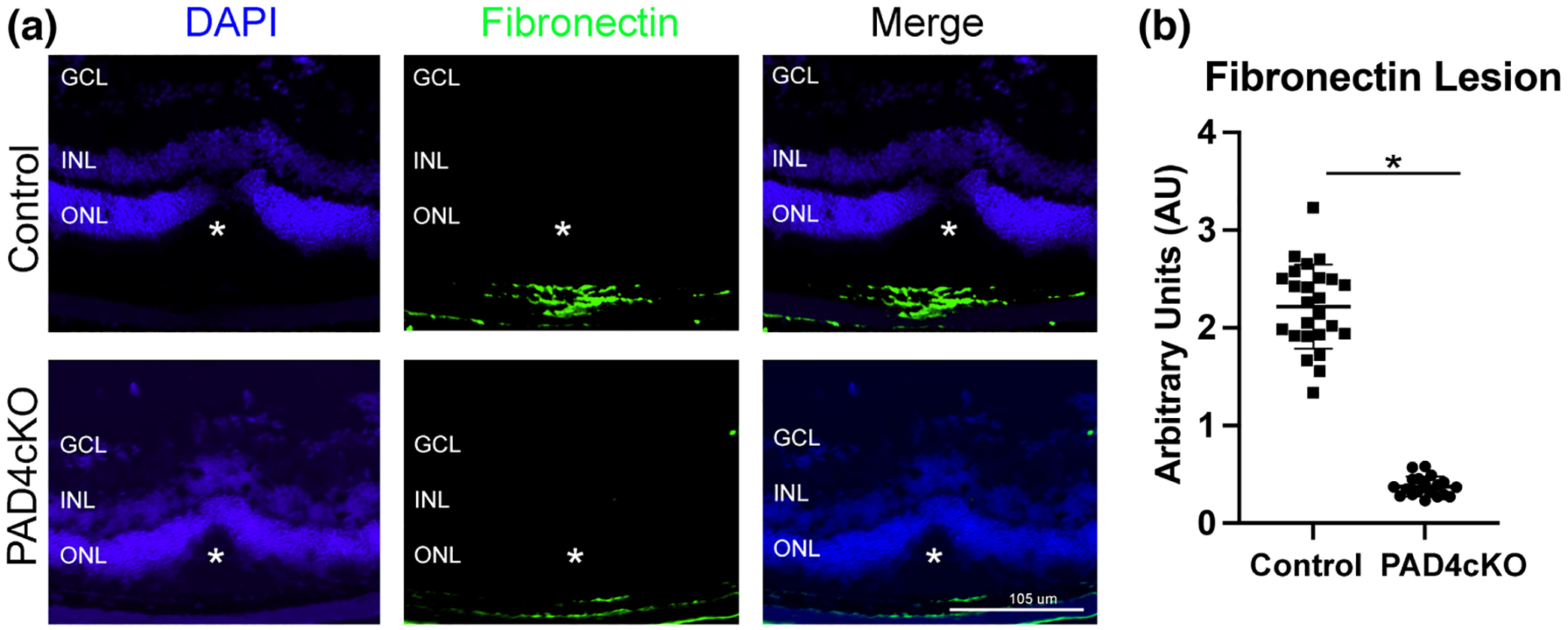
Conditional knockout of PAD4 reduces fibronectin deposition in laser-injured retinas. Representative images of control and PAD4cKO retinas at 30-day post-injury from laser injured mice. (a) Images of control injured and PAD4cKO mice showing staining for fibronectin. Fibronectin deposition in the PAD4cKO lesion site was drastically reduced compared to the control. The asterisks (a) mark the position of laser lesion. (b) The area of staining representing fibronectin was quantified using ImageJ software (see also File S2). Statistical analysis using Welch’s two-tailed parametric t test revealed significant differences between control (mean = 2.219 ± 0.4313) and PAD4cKO (mean = 0.3764 ± 0.1005) samples; t27.36 = 20.63, *p < .0001. N = 6 control mice (25 sections) and N = 6 PAD4cKO mice (19 sections) were analyzed. Data are presented as mean ± SD, p < .05 is considered significant. GCL, ganglion cell layer; INL, inner nuclear layer; ONL, outer nuclear layer.
