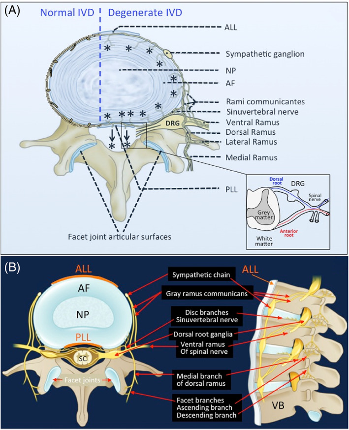
FIGURE 1.

Schematic showing the innervation of the normal and degenerate intervertebral disc (IVD) (A). The normal IVD is the largest predominately aneural structure in the human body where nerves are confined to the outer annular lamellae. With IVD degeneration and depletion of space‐filling aggrecan from the IVD a significant reduction in internal hydrostatic pressure in the IVD and the production of inflammatory mediators and neurotrophic factors provides conditions that are conducive to the ingrowth of nociceptive nerves (*) from the sinu‐intervertebral nerve into the AF of the IVD and significant increases of peripheral mechanoreceptors in the IVD. Thus, there is an increased perception of pain in the mechanically incompetent degenerate IVD. Peripheral IVD neural structures such as the sympathetic ganglion and dorsal root ganglion and the rami communicantes are highly innervated structures. The DRG communicates with the sensory dorsal horn of the spinal cord. There are also connections to the dural nerve plexus (arrows, *) from the sinu‐vertebral nerve. The facet joint capsule and associated subchondral bone and vertebral bodies adjacent to the IVDs and CEPs are all innervated. The spinal nerve has dorsal and anterior connections to the spinal cord (inset). Neural organization of a lumbar spinal segment (B). AF, annulus fibrosus; ALL, anterior longitudinal ligament; DRG, dorsal root ganglion; NP, nucleus pulposus; PLL, posterior longitudinal ligament; SC, spinal cord. Figure modified from Kallewaard et al. 53 with permission
