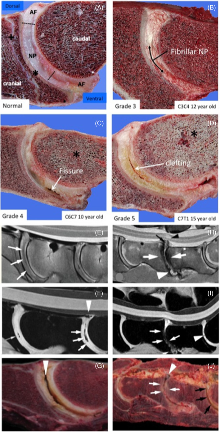
FIGURE 3.

Vertical sections through equine spinal segments showing the characteristic ball and socket appearance of an equine IVD and characteristic tears and lesions that occur with IVDD (A–D). MRI images of equine spinal segments showing reduced MRI signal in the IVD with the onset of IVDD (E, H, F, I) correlating with lesions evident on gross examination of spinal segments (G, J). IVD features are annotated and degenerative clefts and fissures indicated with arrows and decreased bone density changes with an asterisk in (A–D). Images A–D reproduced from 13 under Open Access Attribution‐noncommercial 4.0 International (CC BY‐NC 4.0) license. Diffuse hypo intense regions and signal voids and diffuse hypointense areas are noted throughout the intervertebral discs with loss of definition of the nucleus (white arrows); B, sagittal water selective cartilage image; noted clefts (white arrows) and regions of the dorsal longitudinal ligament of C6‐C7 (white arrow head); and diffuse hypointense areas were noted throughout the IVDs with loss of definition of the nucleus (white arrows); Clefts and loss of NP definition are indicated with white arrows and defects in the longitudinal ligament attachments with arrow heads (E–J). A large central cleft in a C6C7 IVD is indicated by an arrowhead in (G). Severe remodeling of CEPs and discolouration of protruding dorsal IVDs (white arrows) in the C6C7 IVD (J). The C7S1 IVD displays severe cleft formation and yellow discolouration (black arrows)
