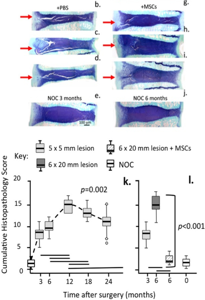
FIGURE 4.

Depiction of how a large 6 × 20 mm outer anterolateral annular lesion (red arrow) destabilizes the ovine IVD and induces disc degeneration with propagation of the lesion into the IVD (b, c, d) and demonstration of the utility of bone marrow derived stromal stem cells for the regeneration of lesion affected IVDs (g, h, i). Toluidine blue‐fast green stained vertical IVD sections. Non‐operated control IVDs (e, j), freshly made lesion in a cadaver IVD (f). Disc degeneration was induced for 4 (b, c, g, h) or 12 weeks (d, i) then injected with PBS (b, c, d) carrier or MSCs (g, h, i) and recovery allowed to proceed for 8 (g, h) or 22 weeks (i). Annular lesions severely reduced the disc heights in the degenerate IVDs (b, c, d) but disc heights were recovered to ~95% of normal values in MSC treated IVDs (g, h, i) where repair of the IVD lesion occurred. In the corresponding PBS carrier treated discs (b, c, d) lesion development was extensive. Histopathological scoring of IVDs 179 in which degeneration had been induced by a 5 × 5 (k) or 6 × 20 mm lesion (l) based on IVD structure, proteoglycan content, disc height, lesion progression, cellular infiltration showed a steady increase in the cumulative degenerate histopathology index in the 5 × 5 mm lesion over 24 months (k) and development of a similar histopathology score by 6 months in the 6 × 20 mm lesion (l) however the administration of MSCs reduced this degeneracy index to levels similar to those evident in non‐operated control IVDs. Figure modified by Open Access under an Attribution 4.0 International CC BY 4.0 license from 162 , 179
