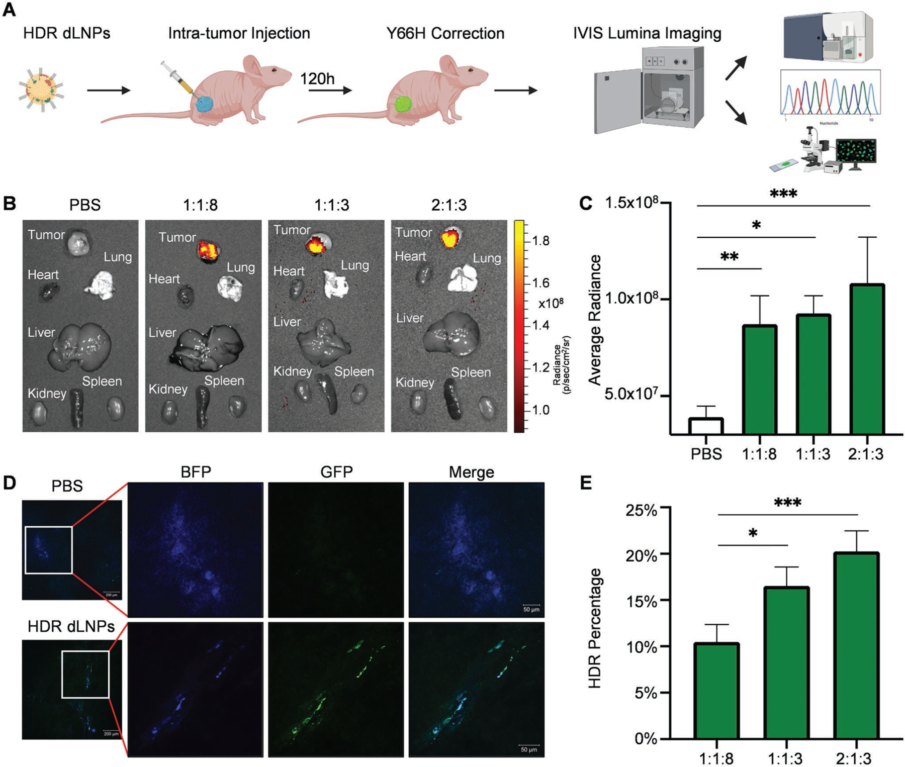
Figure 4.

HDR gene correction was achieved in vivo. A) Subcutaneous xenograft HEK293 (Y66H) tumors were resected 5 days after injection of 4A3-SC8 HDR dLNPs for analysis by IVIS Lumina imaging, flow cytometry, confocal microscopy, and DNA sequencing. B) IVIS Lumina imaging of tumors and organs revealed bright GFP signal in the tumors injected with 1:1:8, 1:1:3, and 2:1:3 HDR dLNPs and no detectable GFP signal in the tumors injected with PBS (N = 3). C) Average radiance of tumors in each HDR dLNP group was quantified revealing a significant difference between all three HDR dLNP and PBS injected tumors (for 1:1:8 p = 0.0060, for 1:1:3 p = 0.0106, for 2:1:3 p = 0.0008; statistical analysis was performed using one-way ANOVA with Tukey’s multiple comparisons test; N = 3). D) Tumors were frozen following resection and sectioned at a thickness of 7 mm for confocal imaging wherein GFP signal was clearly visible in tumors injected with HDR dLNPs. E) gDNA was extracted from whole tumors and then sequenced to obtain HDR correction percentage. Analysis via TIDER revealed average HDR-mediated correction rates of 10.475% for 1:1:8 HDR dLNPs, 16.533% for 1:1:3 HDR dLNPs, and 20.325% for 2:1:3 HDR dLNPs (Statistical analysis was performed using one-way ANOVA with Tukey’s multiple comparisons test, N = 4 with exception of 1:1:3 where N = 3).
