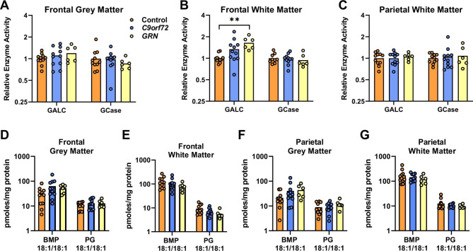
Fig. 5.
Increased GALC activity in frontal white matter of FTD-GRN cases. (A-C) GALC and GCase enzyme activity in frontal white matter (A), parietal white matter (B) and frontal grey matter (C) of control (n = 11), FTD-C9orf72 (n = 11), and FTD-GRN (n = 6) cases. Data is normalised to the mean of the control group. (D-G) Targeted lipidomic analysis of 18:1/18:1 BMP and 18:1/18:1 PG in frontal grey matter (D), frontal white matter (E), parietal grey matter (F), parietal white matter (G). Groups were compared by one-way ANOVA with Tukey’s post-test: **p < 0.01

