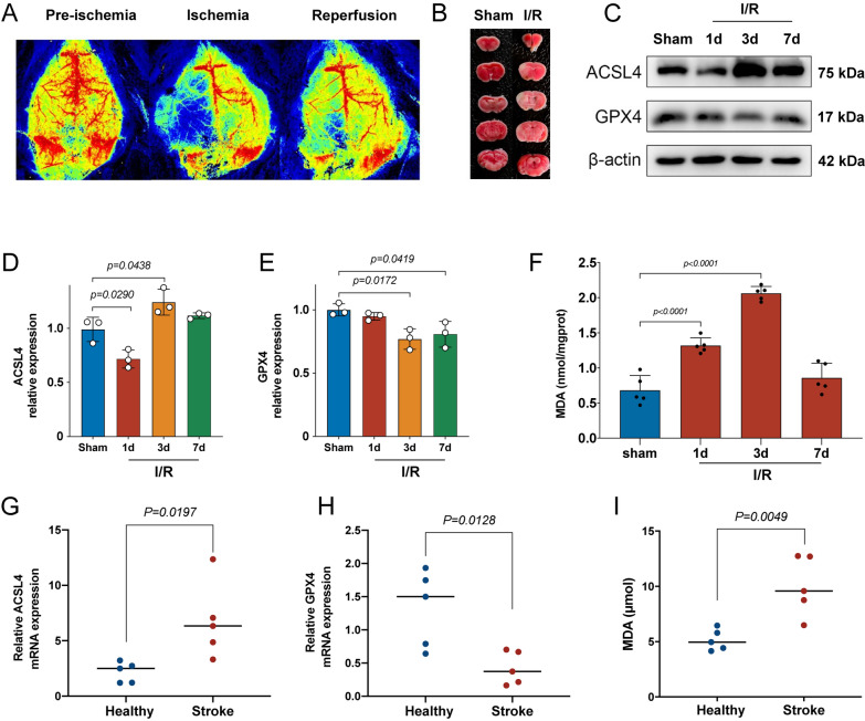
Fig. 3.
Ferroptosis occurred in ischemic stroke mice and patients. A Representative photomicrographs of laser speckle imaging for ischemic stroke mice. B Representative photomicrographs of brain coronal sections stained with triphenyl tetrazolium chloride (TTC) staining for ischemic mice at 24 h after MCAO. C Western blot analysis of ferroptosis markers ACSL4 and GPX4 at different time points after MCAO in mice, n = 3. D Quantification of the protein expression of ACSL4, n = 3. E Quantification of the protein expression of GPX4, n = 3. F Quantification of the malondialdehyde (MDA) at different time points after MCAO in mice, n = 5. G, H The mRNA expression levels of ACSL4 and GPX4 were detected by RT-qPCR in the serum from healthy volunteers and ischemic stroke patients, n = 5. I The amount of MDA detected in the serum from healthy volunteers and ischemic stroke patients, n = 5

