Fig. 5.
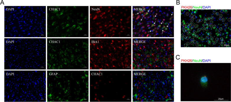
CHAC1 was up-regulated in mouse neurons at 3d after MCAO and ADSC-Exo were taken up by neurons in vivo and in vitro. A The immunofluorescence showed the co-localization of NeuN with CHAC1 obviously, while no co-localization of CHAC1 with IBA1 or GFAP was identified. White arrows indicated the co-localization of NeuN with CHAC1. Scale bars = 20 μm. B Immunofluorescence imaging showed the uptake of ADSC-Exo by neurons in vivo. PKH26 labeled ADSC-Exo (Red) were intranasally administered to the mouse at 24 h after MCAO. The internalization of ADSC-Exo by NeuN + neuron (Green) was detected 6 h after the administration. The white circle indicated ADSC-Exo internalized by neuron. Scale bar = 50 μm. C PKH‐26 labeled ADSC-Exo (Red) were taken up by NeuN + N2a cells (Green). PKH‐26 labeled ADSC-Exo were incubated with N2a cells for 6 h. Scale bar = 20 μm

