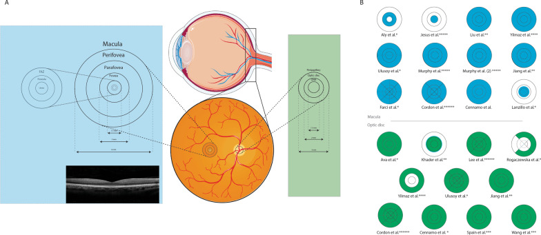
Fig. 1.
Macula and optic disc regions of each included study which were measured for vascular, perfusion, and flow density. A Illustration of retinal fundus and location of the macula and optic disc segments. Foveal avascular zone (FAZ) parts are also displayed in the left circle. B Fields analyzed in each study. Blue shows sections of macula and green shows sections of the optic disc. * indicates studies using an Optovue machine, ** Indicates studies using a Zeiss machine. *** indicates studies using a prototype Axsun SS-OCT machine, **** indicates studies using a Nidek machine, ***** indicates studies using a Heidelberg machine, and ****** indicates studies using a Topcon machine. Note: Parts of the figure were drawn using pictures from Servier Medical Art. Servier Medical Art by Servier is licensed under a Creative Commons Attribution 3.0 Unported License (https://creativecommons.org/licenses/by/3.0/)

