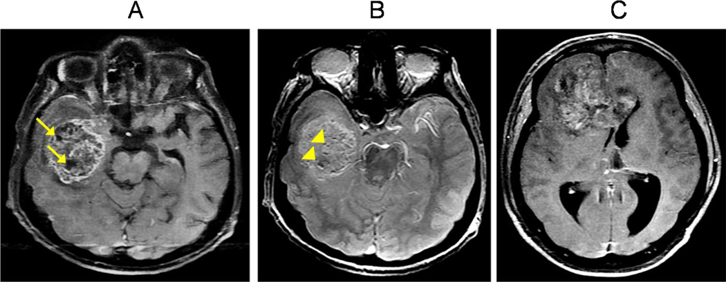
Figure 1. Classic GBM appearances.
(A) T1W contrast-enhanced axial sequence shows an infiltrative mass centered at the right temporal white matter with heterogeneous enhancement and pockets of necrosis (arrows). (B) T2* axial sequence demonstrates multiple susceptibility foci within the mass, suggestive of microhemorrhages (arrowheads), commonly seen with GBMs. (C) T1W contrast-enhanced axial sequence shows a heterogeneously enhancing mass which involves both frontal lobes by crossing via the white matter tracts of the corpus callosum. This “butterfly glioma” pattern is observed in many GBMs.

