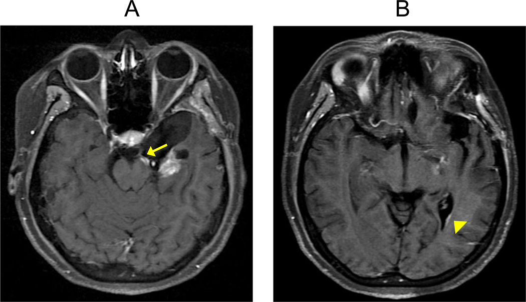
Figure 2. Routes of GBM tumor spread.
(A) Contrast-enhanced T1W axial sequence in a patient with GB which shows enhancement following the cisternal segment of the left oculomotor nerve/CNIII (arrow). The image suggests cerebrospinal fluid (CSF) tumor spread with cranial nerve involvement. (B) Contrast-enhancement T1W axial sequence in the same GBM patient demonstrates linear enhancement along the surface of the left lateral ventricle (arrowhead), suggestive of ependymal tumor spread.

