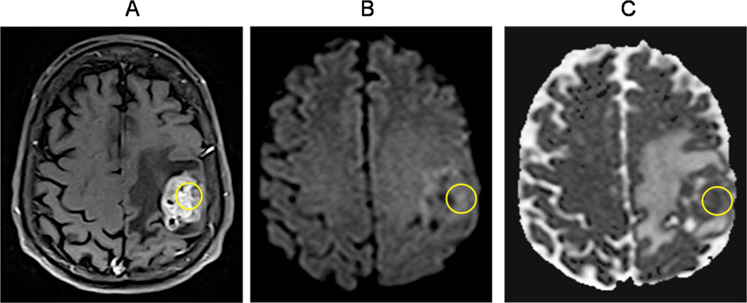
Figure 4. Diffusion weighted imaging (DWI).
(A) Contrast-enhanced T1W axial sequence showing a left frontoparietal necrotic mass in the vicinity of the central sulcus, consistent with a GBM (circle). (B–C) Diffusion weighted image/DWI shows increased signal intensity with corresponding decreased signal intensity on the apparent diffusion coefficient/ADC map within areas of enhancing tumor seen in A (circles). This restricted diffusion pattern is seen in highly cellular tumors such as GBM.

