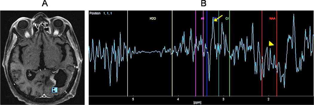
Figure 6. GBM status after post-surgical resection and chemoradiation.
(A) Proton magnetic resonance spectroscopy (H-MRS) was performed using a time of echo (TE) of 136 with single voxel measuring 10×10×10mm placed within a region of focal nodular enhancement at the posterior margin of resection. (B) The resulted spectrum is abnormal with a dominant Choline (Cho) peak (arrow) and a decreasing in the NAA (arrowhead) almost to the level of baseline. Cho/Cr ratio was elevated at 3.1. These findings are highly suggestive of recurrent tumor. These H-MRS images can help to differentiate recurrent tumor from radiation necrosis, which typically causes a decreasing in all peaks without elevation of the Choline peak.

