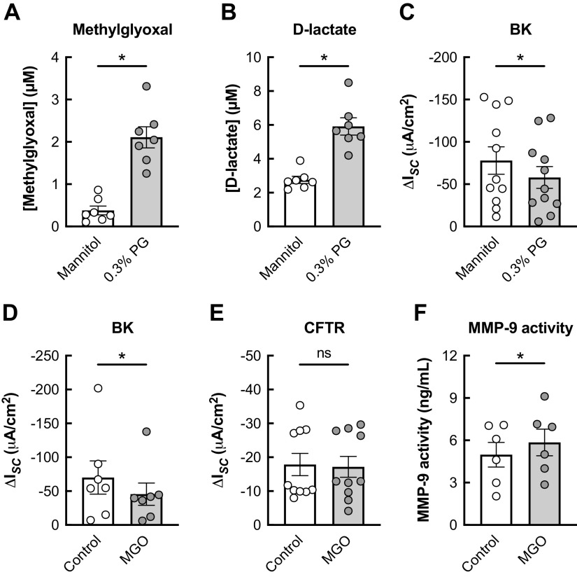
Figure 6.
Basolateral PG inhibits BK channel function in HBECs in vitro. A and B: basolateral levels of methylglyoxal (A) and d-lactate (B) are significantly increased in HBECs 24 h after exposure to basolateral 0.3% PG compared with 0.74% mannitol (isosmotic) control. n = 7 lungs. C: 24-h exposure of HBECs to 0.3% PG causes a significant reduction in BK activity compared with mannitol control. n = 11 from 7 lungs. D: 24-h exposure of HBECs to basolateral MGO (1 µM) causes a significant decrease in BK function. n = 7 lungs. E: basolateral MGO does not significantly change CFTR conductance compared with controls after 24 h. n = 10 from 6 lungs. F: 24-h exposure of HBECs to basolateral MGO causes a significant increase in MMP-9 activity measured in the basolateral media. n = 6 from 3 lungs. Data are presented as means ± SE. *P < 0.05, ns = not significant. Data were analyzed by two-tailed t test (A–D and F) or two-tailed Wilcoxon test (E) after assessing normality by Shapiro-Wilk. BK, large conductance, Ca2+-activated, and voltage-dependent K+; HBECs, human bronchial epithelial cells; MGO, methylglyoxal; PG, propylene glycol.

