. 2022 Jul 14;41(2):422–433. doi: 10.5534/wjmh.220009

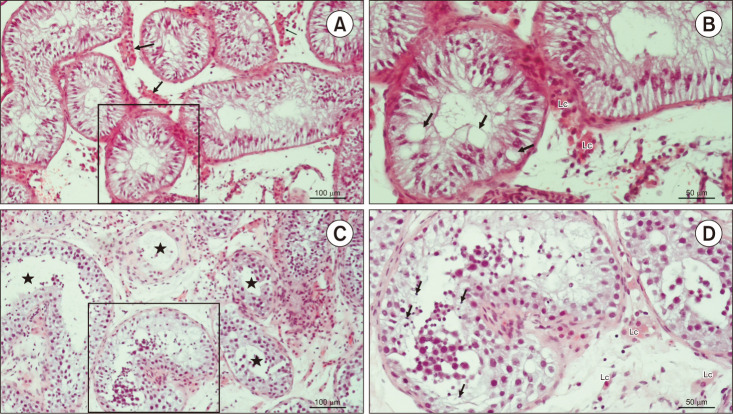
Fig. 2. Testicular histology of patients code 160585017 and 141879319. (A) Testicular biopsy from an azoospermic patient. Seminiferous tubules display narrow lumen and are lined with Sertoli cells exclusively. Lc clusters are seen between the seminiferous tubules (→) (H&E, ×200, scale=100 µm). Patient 160585017. (B) Detail from panel A. Due to the depletion of spermatogenic cells, Sertoli cells display giant vacuoles in their cytoplasm (→) (H&E, ×200, scale=50 µm). Patient 160585017. (C) An azoospermic patient with mixed atrophy of seminiferous tubules. Seminiferous epithelium displays a variable status of spermatogenesis (★) (H&E, ×200, scale=100 µm). Patient 141879319. (D) Detail from panel C. In one of the seminiferous tubules, late spermatids (→) are clearly visible, despite the disorganisation of the seminiferous epithelium (H&E, ×200, scale bar=50 µm). Patient 141879319. Lc: Leydig cells.

Fig. 2