. 2022 Jul 14;41(2):422–433. doi: 10.5534/wjmh.220009

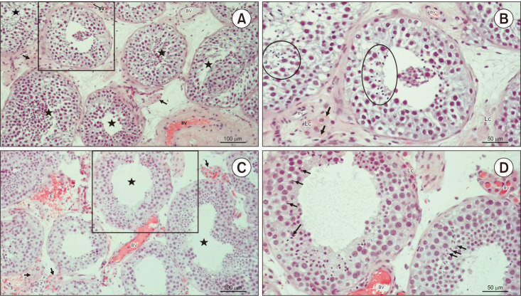
Fig. 3. Testicular histology of patients code 265952375 and 119526696. (A) Testicular biopsy from a patient with hypospermatogenesis. In most tubules, (★) seminiferous epithelium is slightly disorganised but bears late spermatids. Within the loose connective tissue, there are clusters of Lc of moderate size (→) (H&E, ×200, scale bar=100 µm). Patient 265952375. (B) Detail from panel A. Despite changed stratification of spermatogenic cells, seminiferous tubules preserved the capacity to produce late spermatids (circled areas). The interstitial compartment bears small BV and Lc. Some Lc are rich in Reinke’s crystals (→) (H&E, ×200, scale bar=50 µm). Patient 265952375. (C) Testicular parenchyma sample from an azoospermic man. Seminiferous tubules display heterogeneous morphology of spermatogenesis (hypospermatogenesis, maturation arrest), including some tubules rich in late spermatids (★). Clusters of Lc (→) have an abundant network of accompanying capillaries. As a consequence of surgery, extravasated erythrocytes (E) are frequently encountered in the bioptic material (H&E, ×200, scale bar=100 µm). Patient 119526696. (D) Detail of panel C. These two seminiferous tubules have maintained a regular diameter and morphology of spermatogenesis. Some late spermatids (→) are in the final stage of spermiogenesis and will be released as spermatozoa in the lumen. Peritubular Lc are found adjacent to the lamina propria of the tubule. The interstitium is rich in BV (H&E, ×200, scale=50 µm). Patient 119526696. Lc: Leydig cells, BV: blood vessels.

Fig. 3