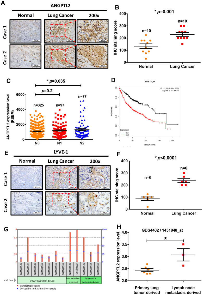
Figure 2.
Levels of ANGPTL2 expression correlate with clinicopathologic features of lung adenocarcinoma tissue infiltrated by lymphatic vessels. (A, B) ANGPTL2 expression in human lung cancer tissue and adjacent normal tissue samples was analyzed by IHC staining. (C) The association between ANGPTL2 expression and regional lymph node metastasis was analyzed in samples from the TCGA database. (D) Associations between ANGPTL2 expression and overall survival rates of lung cancer patients were analyzed using the Kaplan-Meier Plotter database. (E, F) LYVE-1 expression in human lung cancer tissue and adjacent normal tissue samples was analyzed by IHC staining. (G, H) Data obtained from the GEO database (GDS4402/1431848_at) were analyzed for ANGPTL2 expression in primary lung tumor tissue and lung cancer with lymph node metastasis tissue samples. *p < 0.05 compared with normal tissue.

