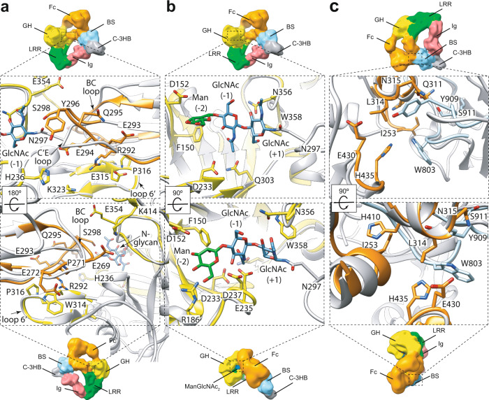
Fig. 2. The Fc binding site of EndoS.
In order to have insights on the possible location of the side chain residues that could be involved in the enzyme-substrate interactions, we locally compared/superimposed the high-resolution EndoS (PDB code 6EN3; yellow) and Fc region (PDB code 2DTS; orange) crystal structures with our EndoSE235A-Fc cryoEM structure (gray; Methods section). Therefore, we visualize potential side chain interactions between residues that are within interaction distance based on flexible fitting of the superimposed crystal structures. Two views of the interaction interface between the GH domain of EndoS (yellow, PDB code 6EN3) and FcP1 of the Fc region (orange, PDB code 2DTS) (a), the N-glycan (blue and green) of the Fc region and the active site of EndoS (yellow, PDB code 4NUY) (b) and the Cγ2-Cγ3 joint region of the Fc (orange, PDB code 2DTS) and the β-sandwich domain of EndoS (blue, PDB code 4NUY) (c).

