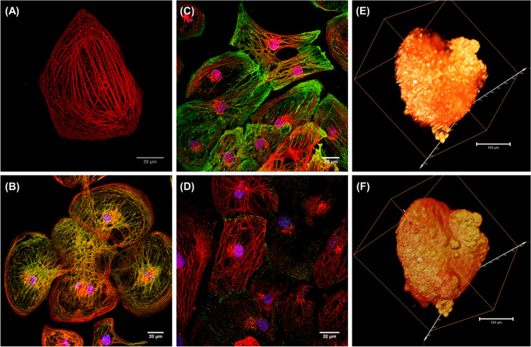
Fig. 3.
Immunofluorescence images of MYH6-mScarlet reporter CMs expressing cardiac structural markers. Confocal image of fixed reporter CMs showing the myofibril striations (A). Reporter CMs immunostained with cardiac troponin T (cTnT) green (B), myosin-binding protein (MYBPC) green (C), and gap junction connexin 43 (Cx43) green (D). Nuclei were counterstained with DAPI (blue) in all immunostained images. Scale bars are 20 μm. Single/selective plane illumination microscopy (SPIM) optical 3D stack of multi-focal mScarlet fluorescence showing the spatial distribution (E) and topography of cardiomyocytes in an aggregate within cluster (F)

