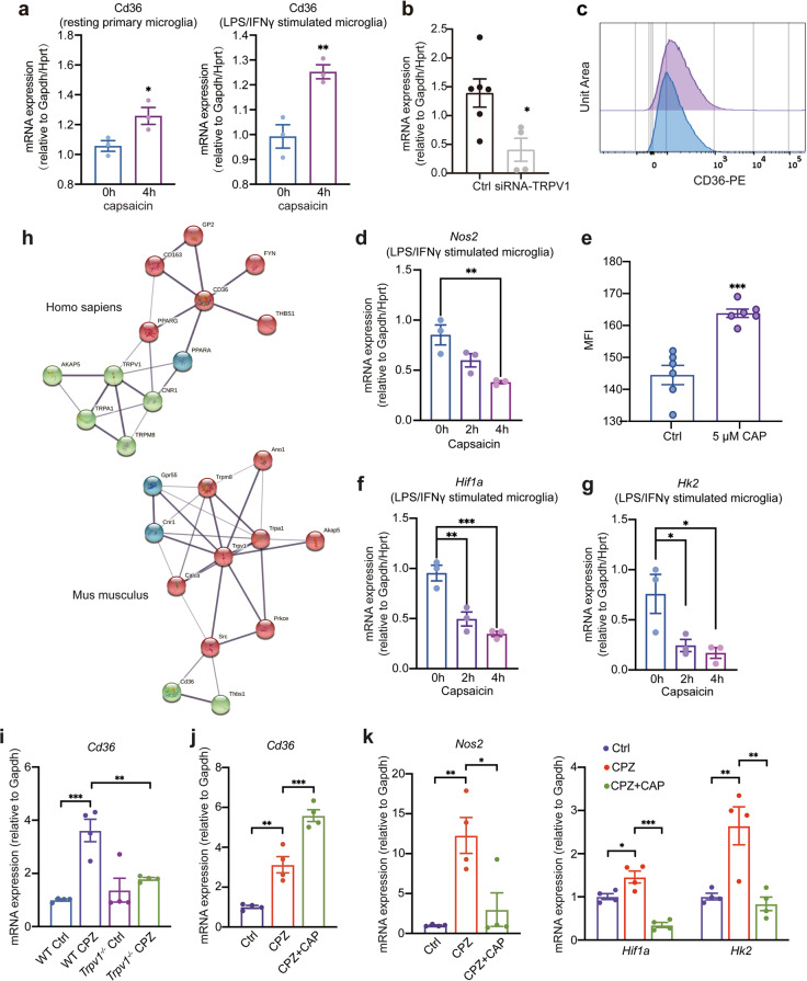
Fig. 7. The scavenger receptor CD36 is involved during TRPV1 activation.
a mRNA expression of Cd36 (normalized to Gapdh and Hprt) of resting and LPS/IFNγ-stimulated primary microglia treated with capsaicin for 4 h (mean ± SEM; n = 3; *P < 0.05, **P < 0.01 by unpaired two-tailed t-test). b mRNA expression of Cd36 (normalized to Gapdh and Hprt) of control microglia or siRNA-TRPV1 treated microglia after 24 h (mean ± SEM; n = 4 or 6; *P < 0.05 by unpaired two-tailed t-test). c, e Flow cytometry analysis of CD36 expression in untreated BV2 microglia or treated with 5 μM CAP for 24 h (mean ± SEM; n = 6; ***P < 0.001 by unpaired two-tailed t-test). mRNA expression of Nos2 (d), Hif1a (f), and Hk2 (g) in LPS/IFNγ-stimulated microglia treated with capsaicin for indicated hours (mean ± SEM; n = 3; *P < 0.05, **P < 0.01, ***P < 0.001 by one-way ANOVA with Dunnett’s multiple comparisons test). h STRING analysis showing the close interaction between TRPV1 and CD36 in Homo sapiens and Mus musculus. i Gene expression analysis of Cd36 in the corpus callosum of WT and Trpv1−/− mice (mean ± SEM; n = 4 mice for each group; **P < 0.01, ***P < 0.001 by two-way ANOVA with Sidak’s multiple comparisons test). j, k Expression of Cd36, Nos2, Hif1a and Hk2 was assessed by RT-PCR in the corpus callosum at the end of capsaicin treatment (mean ± SEM; n = 4; *P < 0.05, **P < 0.01, ***P < 0.001 by one-way ANOVA with Tukey’s multiple comparisons test).

