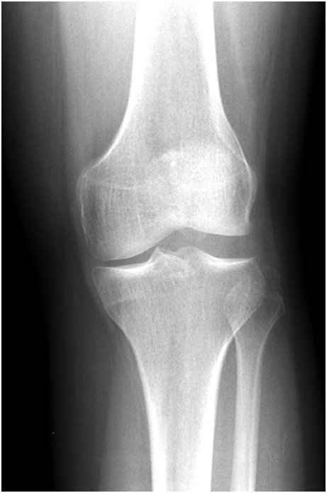
Fig. 1.

Anteroposterior radiograph of the left knee of a skeletally mature patient demonstrating all four radiographic findings of DLM: squaring of the lateral femoral condyle, concavity of the lateral tibial plateau, increased lateral joint space, and hypoplasia of the lateral tibial spine
