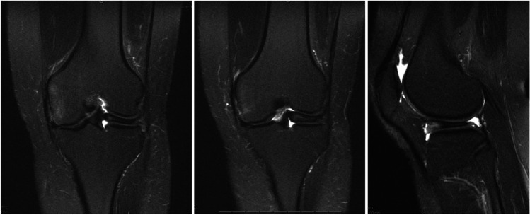
Fig. 2.
MRI T2 coronal × 2 and sagittal images demonstrating a skeletally mature patient with DLM. Increased T2 signal seen within the meniscal tissue as a horizontal strip represents intrasubstance degeneration with myxoid tissue. This is seen most clearly on T2 images due to increased water content due to structural dysregulation within the meniscal tissue. Hypoplasia of the lateral femoral condyle is also appreciated

