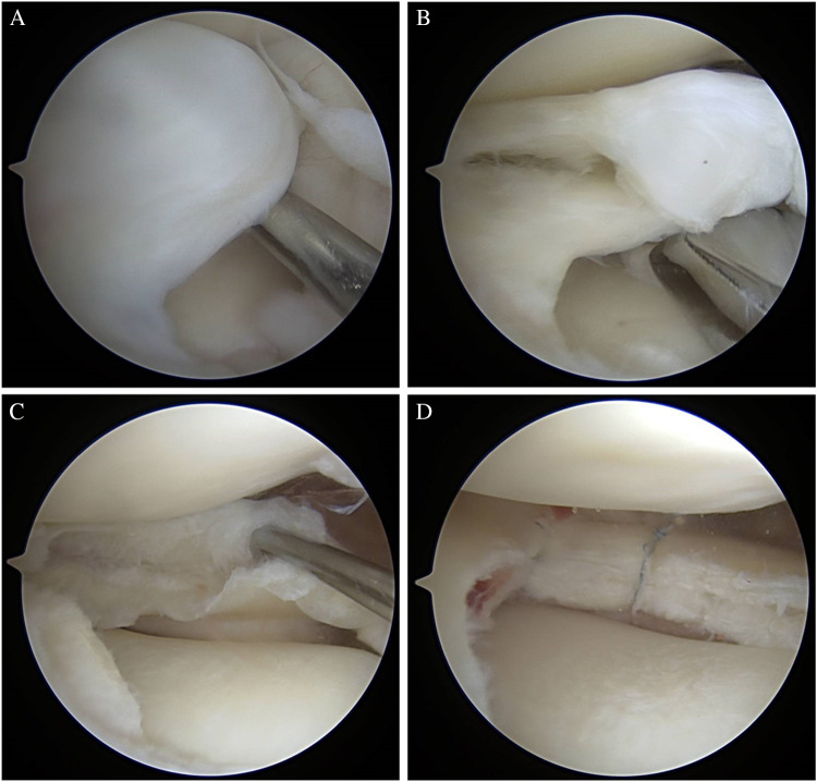
Fig. 5.
A–D: arthroscopic images of a right knee. (A) DLM with full coverage of the lateral tibial plateau. (B–C) ID revealed as sauceration is performed. (D) all-inside capsular-based anchors being placed with similar compression of leaflets to Fig. 4 images. The difference in this case is the repair technique with capsular-based repair stitches in this figure and all-meniscal suture without capsular inclusion in Fig. 4

