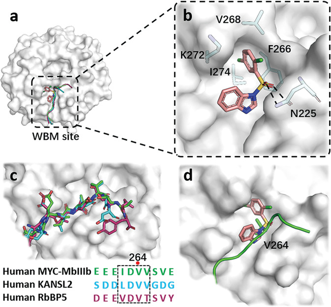
Fig. 4. X-ray crystal structure of WDR5 in complexed with compound 19.
a A surface representation of compound 19 and MYC-MbIIIb, KANSL2, RbBP5 peptides interactions with WDR5 at WBM site. b Detailed interactions of compound 19 with pivotal residues of WDR5 is shown as sticks and hydrogen bonds are shown as black dash lines. c Superimposition of the conserved MbIIIb, KANSL2 and RbBP5 in complex with WDR5, and the sequence alignment of the peptides. d Superimposed binding modes of compound 19 and MYC-MbIIIb with WDR5 at WBM site.

