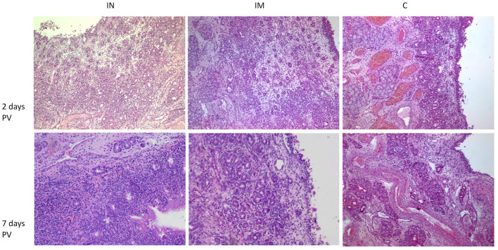
Figure 5.
Histological features of nasal mucosa sections in the IN-vaccinated group, IM-vaccinated group and C group at 2 days and 7 days PV. H&E (100× magnification). IN group: at 2 days PV, epithelium, superficial and deep corium are diffusely infiltrated by lymphocytes, plasma cells and macrophages with superficial edema; at 7 days PV, within superficial corium, a diffuse and dense inflammatory infiltrate composed predominantly by numerous lymphocytes and plasma cells is present. Numerous intraepithelial lymphocytes are visible within epithelium. IM group: at 2 days PV, deep corium is multifocally infiltrated mainly by lymphocytes and plasma cells, while superficial corium is edematous; at 7 days PV, within superficial corium, a periglandular lymphoplasmacytic infiltrate is present; the epithelium is focally eroded. C group: at 2 days PV, within superficial corium, a multifocal lymphoplasmacytic infiltrate is present; at 7 days PV within superficial corium, a low number of lymphocytes and plasma cells surround glands and capillaries. IN, intranasally; IM, intramuscularly; C, control; PV, post-vaccination; H&E, hematoxylin and eosin.

