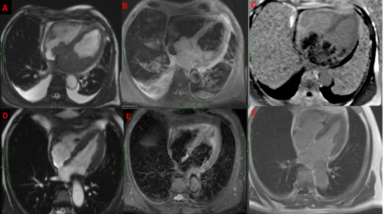
Figure 2.
CMR showing a bulky mass extending throughout the left atrium, infiltrating the interatrial septum and right atrium, on cine imaging sequence (A), slightly hyperintense on fat-saturated T2-weighted imaging (B) with heterogeneous enhancement on late gadolinium enhancement sequences (C) with complete disappearance after 6 cycles of R-CHOP in the same sequences (D–F).

