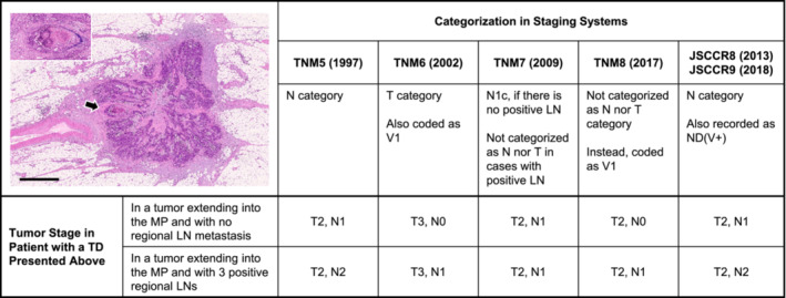
FIGURE 4.

Stage migration caused by different categorizations of a tumor deposit (TD) depending on staging systems. The picture in the upper‐left panel indicates a peritumoral TD with a diameter of approximately 3.5 mm with an irregular contour and an identifiable vascular structure. Under TNM5, this nodule is classified as an LN because it is >3 mm in diameter. On the contrary, this nodule is considered a lesion of the T category because of its contour and is also coded as venous invasion under TNM6. The category N1c is used for this nodule in the absence of regional LN metastasis under TNM7, whereas under TNM8, the tumor stage does not change by this nodule which is regarded as venous invasion because the vascular structure is evident (arrow). Since 2013, this nodule has been invariably treated the same as LN metastasis to derive the final N stage in Japan. Picture, hematoxylin and eosin staining; bar, 1 mm. The inset illustrates the magnification of the part of the nodule that is indicated with an arrow (Victoria blue–hematoxylin and eosin staining). LN, Lymph node; MP, Muscularis propria; TD: Tumor deposit
