Fig. 5.

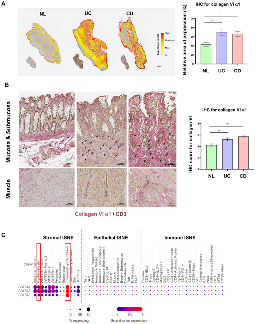
The expression of collagen VI is elevated in inflamed colon tissue in inflammatory bowel disease.
(A) Collagen VI α1 immunohistochemistry of full thickness intestinal tissue sections in ulcerative colitis (UC) and Crohn’s disease (CD), compared to normal control (NL). Automatic quantification by HALO software showed significantly higher expression in UC and CD compared to NL (n = 5, t test). (B) Collagen VI α1 expression on immunohistochemistry was elevated in the mucosa, submucosa and muscle layers in UC and CD, compared to NL. Under high magnification CD3 positive immune cells (brown) were found encased by and in close proximity to collagen VI α1 (red) in the submucosa of inflammatory bowel disease (IBD) tissues (black arrows). The increase in collagen VI α1 in intestinal tissues was confirmed by blinded scoring of full thickness colonic sections using a prespecified scoring system (n = 12, t test). (C) Open access single cell dataset [29] revealed that COL6A1, COL6A2 and COL6A3 were mainly expressed in stromal cells in UC, especially in inflammatory fibroblasts, but not in epithelial and immune cells in UC. *, p < 0.05, **, p < 0.01, ***, p < 0.001.
