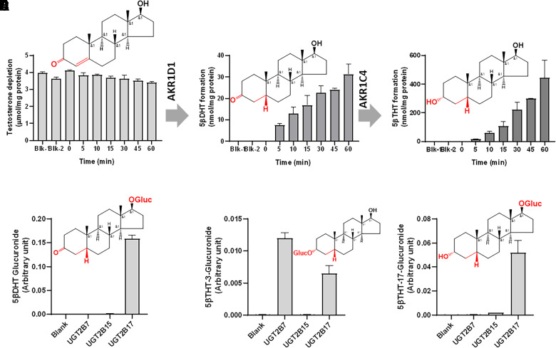
Fig. 3.
In vitro testosterone metabolism by aldoketoreductases in liver cytosol samples from the UGT2B17 deletion subjects (A–C) and in the recombinant UGTs (D–F). Testosterone to 5β-DHT and 5β-THT conversion with time by AKR1D1 and AKR1C4 in the human liver cytosol obtained from the UGT2B17 deletion subjects (A–C). Blk-1 and Blk-2 samples do not conatin NADPH and the enzyme, respectively. In vitro glucuronidation of 5β-DHT and 5β-THT using the recombinant human UGT2B enzymes (D–F). Structures of testosterone, 5β-DHT, 3α,5β-THT, 5β-DHT-glucuronide, 3α,5β-THT-3-glucuronide, and 3α,5β-THT-17-glucuronide are shown in (A–F), respectively.

