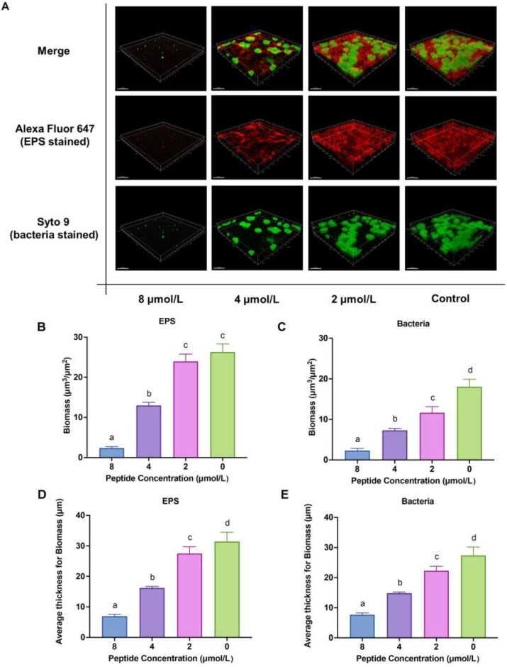
Figure 3.
The three-dimensional structure of Streptococcus mutans biofilms under the LF-1 treatment for 24 h is observed by the CLSM, where EPS is stained red with Alexa Fluor 647 and bacteria are stained green with Syto 9. (A) Biomass (B,C) and average thickness (D,E) for EPS and bacteria are calculated according to five randomly selected images from the red and green channels, respectively. Data are presented as the mean ± standard deviation, and columns labelled with different superscript letters denote significant statistical differences in one group (one-way analysis of variance; p < 0.05). EPS: exopolysaccharide; CLSM: confocal laser scanning microscopy.

