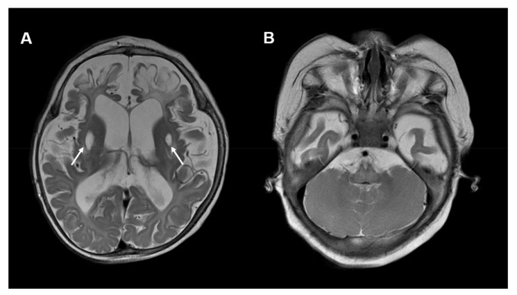
Figure 4.
Neuroimaging in COQ7 deficiency: (A) Brain MRI, T2-weighted, axial images of a 10-month-old boy with COQ7 deficiency. The MRI shows global brain atrophy and areas of encephalomalacia in bilateral frontal lobes. In addition, symmetric cystic changes within the putamen are visible (white arrows). (B) No cerebellar abnormalities are visible. Other MRI images of this individual were published previously [41].

