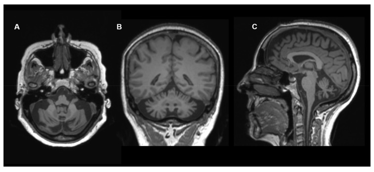
Figure 5.
Neuroimaging in COQ8A deficiency: Brain MRI (T1-weighted images, (A) axial view; (B) coronal view; (C) sagittal view) of a 60-year-old female with COQ8A deficiency. Images show cerebellar atrophy. Other MRI images of this individual were published previously [45].

