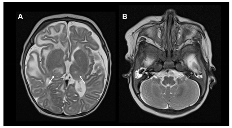
Figure 6.
Neuroimaging in HPDL deficiency: (A) Brain MRI, T2-weighted images of a 3-month-old girl with HPDL deficiency. Images show subcortical T2-hyperintensities, mainly affecting the right frontotemporal regions. Moreover, asymmetrical signal abnormalities of the thalami are visible (white arrows). (B) No cerebellar abnormalities are visible.

