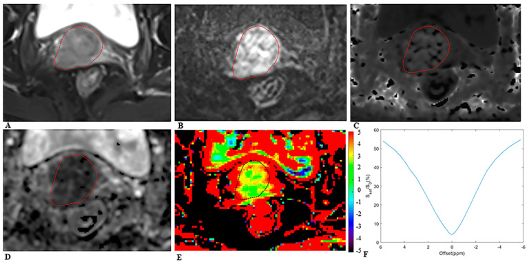
Figure 2.
MRI scans in a 64−year−old woman with cervical cancer and postoperative pathology revealed LNM. Axial (A) T2−weighted image illustrates an exophytic tumor on the cervix wall. A diffusion−weighted image (B) with b = 1000 s/mm2 shows a high−signal−intensity tumor. Mean kurtosis (MK) map (C) and mean diffusivity (MD) map (D) generated from DKI model. Amide proton transfer−weighted (APTw) image (E) and Z−spectrum of the tumor (F); the color bar indicates the APTw value. The mean MK, MD, and APTw values measured by the two radiologists were 1.116, 0.774 × 10⁻³ mm2/s, and 4.5%, respectively.

